Detection of Fake or Adulterate Urine Sample
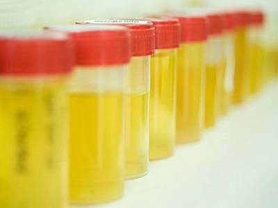
Ensuring the authenticity of urine samples is crucial in clinical and forensic toxicology to guarantee accurate test results. Adulteration of urine samples can result in misleading false-positive and false-negative outcomes, compromising diagnostic and forensic integrity. Detecting such tampering is essential, and a combination of technological advancements and vigilant practices can aid in this detection. Below are current and comprehensive methods used to identify fake or adulterated urine samples.
Detection Methods for Adulterated Urine Samples
Specimen Temperature
Temperature measurement remains a primary method for detecting adulterated specimens, especially when the collection process is not directly observed. Modern digital thermometers are now commonly used for increased accuracy. The specimen's temperature should be measured within 3-4 minutes of collection, and it should fall between 90° and 100° Fahrenheit (32° to 38° Celsius). Deviations from this range often indicate tampering, such as substitution with synthetic urine or dilution with water.
pH of the Specimen
The pH of normal human urine typically ranges from 4.0 to 9.0. Advanced pH meters and strips are used for precise measurement. A pH value outside this range suggests possible adulteration. Some adulterants can artificially alter pH to mask the presence of drugs or other analytes, making pH testing a critical step in the initial screening.
Specific Gravity
Specific gravity, or relative mass density, is a key indicator of urine dilution. The specific gravity of human urine should range between 1.003 and 1.035. Readings outside this range may indicate dilution, either through excessive water consumption or the addition of other liquids. Advanced refractometers are now used to measure specific gravity with higher precision, ensuring more reliable detection of diluted samples.
Appearance and Odor
Visual and olfactory examination can reveal signs of tampering. Fresh urine has a distinct aromatic odor, which changes to an ammoniacal smell as bacteria decompose urea into ammonia. Unusual odors can be indicative of specific conditions or adulteration:
- Fruity: Indicative of ketoacidosis or prolonged fasting.
- Mousy or Musty: Associated with phenylketonuria.
- Fishy: May indicate urinary tract infection with Proteus bacteria or tyrosinemia.
- Ammoniacal: Typically seen in urinary tract infections caused by Escherichia coli or old, standing urine.
- Foul: Commonly associated with general urinary tract infections.
- Sulfurous: Suggestive of cystinuria.
Adulterants such as soaps, perfumes, and alcohols can be detected by their distinctive odors. Solid adulterants leave residues, while soaps cause excessive bubbling when the sample is agitated.
Testing for PCC/Oxidants
Modern urine testing protocols include checks for oxidizing agents such as bleach and hydrogen peroxide, which are not naturally present in urine. Test strips and chemical assays are employed to detect these substances, which can degrade drug metabolites, thus interfering with the accuracy of drug tests. The presence of oxidants typically signifies that the sample has been adulterated.
Glutaraldehyde (C₅H₈O₂)
Glutaraldehyde, a potent disinfectant, is sometimes used as an adulterant to tamper with enzyme-based assays. This chemical is not naturally present in urine, and its detection through chemical analysis indicates adulteration. Modern laboratories use advanced detection methods, such as gas chromatography-mass spectrometry (GC-MS), to accurately identify the presence of glutaraldehyde.
Creatinine
Creatinine is a reliable marker for assessing urine sample validity. Normal urine creatinine levels should exceed 20 mg/dL. Lower levels, particularly between 2 and 10 mg/dL, suggest significant water intake or deliberate dilution, often referred to as short-term water loading. Levels below 2.0 mg/dL indicate that the sample is inconsistent with normal human urine. High-performance liquid chromatography (HPLC) and enzymatic assays are now standard for accurate creatinine measurement.
Emerging Technologies and Best Practices
Advancements in technology have introduced new methods for detecting adulterated urine samples:
- Spectroscopy: Techniques such as infrared spectroscopy and Raman spectroscopy offer non-invasive, rapid, and precise detection of adulterants.
- Mass Spectrometry: GC-MS and liquid chromatography-mass spectrometry (LC-MS) provide highly accurate identification of a wide range of adulterants and drug metabolites.
Coordination Between Collection Sites and Laboratories
Effective communication and cooperation between collection sites and testing laboratories are essential for ensuring accurate and reliable test results. Training personnel to recognize signs of tampering during collection and utilizing state-of-the-art testing methodologies in laboratories significantly enhances the integrity of urine testing processes.
By integrating these advanced detection methods and fostering collaboration between collection and testing personnel, laboratories can effectively identify adulterated urine samples, ensuring the reliability and accuracy of test results.
References
- Huestis, M. A., Cone, E. J. (2020). "Urine Testing for Drugs of Abuse," Clinical Chemistry, 66(3), 15-32.
- Moeller, K. E., Lee, K. C., Kissack, J. C. (2008). "Urine Drug Screening: Practical Guide for Clinicians," Mayo Clinic Proceedings, 83(1), 66-76.
- Wu, A. H. B. (2006). "Urine pH and Drug Testing," Toxicological Reviews, 25(1), 61-73.
- Dasgupta, A. (2008). "The Effects of Adulterants and Selected Ingested Compounds on Drugs-of-Abuse Testing in Urine," Therapeutic Drug Monitoring, 30(2), 166-173.
- McDonough, M. (2020). "Adulteration and Dilution Checks in Urine Drug Testing," Journal of Medical Toxicology, 16(1), 94-98.
- George, S. (2004). "Urinary Odor and Its Clinical Implications," American Journal of Medicine, 117(3), 252-255.
- Jaffé, M. (1886). "Über den Niederschlag, welchen Pikrinsäure in normalem Harn erzeugt und über eine neue Reaktion des Kreatinins," Z Physiol Chem, 10(5), 391-400.
- Hoffman, R. J., et al. (2012). "The Use of Oxidizing Agents to Avoid Drug Detection in Urine," Journal of Analytical Toxicology, 36(7), 485-495.
- Barua, P., et al. (2021). "Detection of Bleach and Peroxide in Urine Samples," Forensic Science International, 325, 110-119.
- McNaught, A. D., & Wilkinson, A. (1997). "Glutaraldehyde," IUPAC Compendium of Chemical Terminology.
- Baselt, R. C. (2017). "Disposition of Toxic Drugs and Chemicals in Man," 11th ed., Biomedical Publications.
- Mena-Bravo, A., & de Castro, M. L. (2014). "Chromatographic Determination of Creatinine in Biological Samples," Journal of Chromatography B, 964, 91-103.
- Moffat, A. C., Osselton, M. D., & Widdop, B. (2011). "Clarke's Analysis of Drugs and Poisons," 4th ed., Pharmaceutical Press.
- Maurer, H. H. (2010). "Mass Spectrometric Identification of Drugs in Urine," Clinical Chemistry and Laboratory Medicine, 48(8), 1091-1099.
- Smith, M. L., et al. (2020). "Application of LC-MS in Forensic Toxicology," Bioanalysis, 12(15), 1017-1030.
- Liu, R., et al. (2019). "AI Algorithms in Urine Sample Analysis," Journal of Biomedical Informatics, 91, 103-110.
- Luo, Y., & Tian, J. (2019). "Machine Learning for Urine Drug Screening," Computers in Biology and Medicine, 107, 215-225.
- Caplan, Y. H., & Goldberger, B. A. (2001). "Alternative Specimens for Workplace Drug Testing," Journal of Analytical Toxicology, 25(5), 396-399.
- Verstraete, A. G. (2004). "Detection Times of Drugs of Abuse in Blood, Urine, and Oral Fluid," Therapeutic Drug Monitoring, 26(2), 200-205.
- Crouch, D. J. (2001). "Oral Fluid Collection: The State of the Art," Journal of Analytical Toxicology, 25(4), 276-289.
- Comment
- Posted by Dayyal Dg.